Wow, what an experience. If you follow this blog, you probably know by now that giving back is very important to me. Throughout my treatment I volunteered for numerous studies and research projects to hopefully make the life of future patients a little easier. So when I heard about Pediatric Preclinical Modelling Program through the ACCESS (Accelerating Childhood Cancer Experience, Science and Survivorship) network, I knew I wanted to be involved right away.
The xenotransplantation program transplants both human cancer cell lines and patient-derived tumour cells into larval zebrafish to better understand cancer progression and to find more personalized therapies. Cells can be tracked in real-time for proliferation, migration and homing to tissue niches. This is groundbreaking and could lead to more personalized treatment plans and better suited drugs to fight childhood cancer.
My role is as a PWLE, or Person With Lived Experience. I help remind the researchers the reason they do what they do and the impact it can have on a patient’s long-term outcome. I also give them the value of a patient’s perspective and how they feel being a part of the program. This includes the emotions they may feel, how and why they would like to be involved in their own treatment, family input, and other perspectives that would otherwise not be considered.
During my introductory meeting with the team, I was invited to the University of Ottawa to visit the Berman Lab. I jumped at this unique opportunity! When we arrived at the lab, the first thing I noticed was the actuator on the door. Big thumbs up for accessibility! We were greeted by Nadine Azzam, the Research Coordinator who brought us up to introduce us to the rest of the team. They were: Jennifer Fiene (Graduate Student), Lissandra Tuzi (Research Technician), Serkan Dogan (Research Technician), Sergey Prykhozhij (Research Associate), Priscilla Fung (Graduate Student), Kim Kobar (Graduate Student), Kevin Ban (Research Technician), Sarada Ketharnathan (Research Associate), Kathleen Connolly (Undergraduate Student), and Maggie Divok (Undergraduate Student).
We were then joined by Jason Berman himself. We had a great discussion about my treatment and how the work they are doing could one day totally change the way people with a similar diagnosis are treated. As I learned about the program from them, they also learned from my experience. It was a beneficial exchange for both parties.
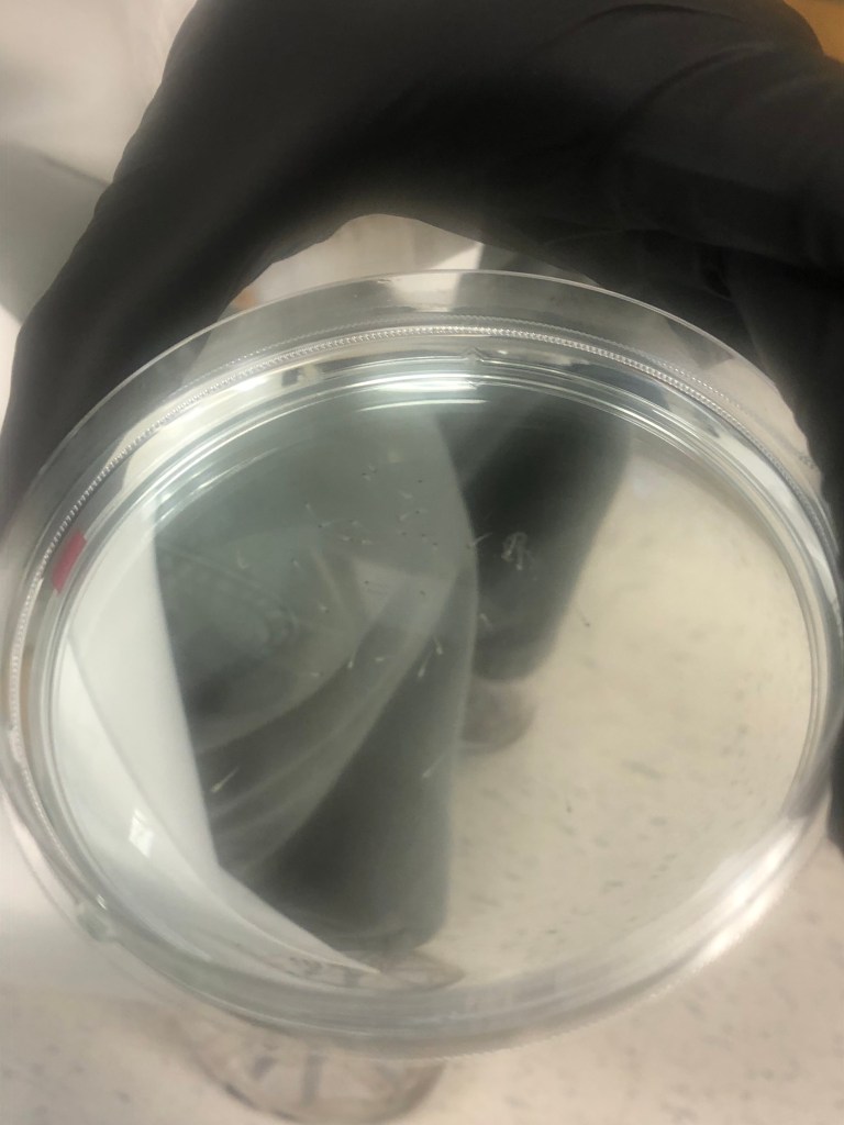
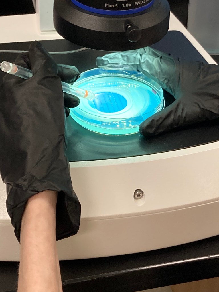
It was then time to start the tour. Our first stop was at the injection phase. This is where they inject the larval zebrafish with the cancer cells in the yolk, the hindbrain, or in the circulation, depending on the cancer type. This is done through a microscope as these are super tiny! A needle smaller than a hair is loaded with the cells, then the operator uses a small foot pedal to push a miniscule puff of air through the needle to inject into the larval. I actually got to try my hand at injecting! Obviously I used dye, not real cancer cells, but I was given the chance to hold the needle and puff the air through to inject. Trust me, this is harder than it looks, and it doesn’t look easy! Definitely no coffee on injection days haha. What I loved was that the air pedal could be moved so you could use any part of your body to press it rather than just your foot, rendering it more accessible for disabled researchers as they could use their elbows or their other hand.
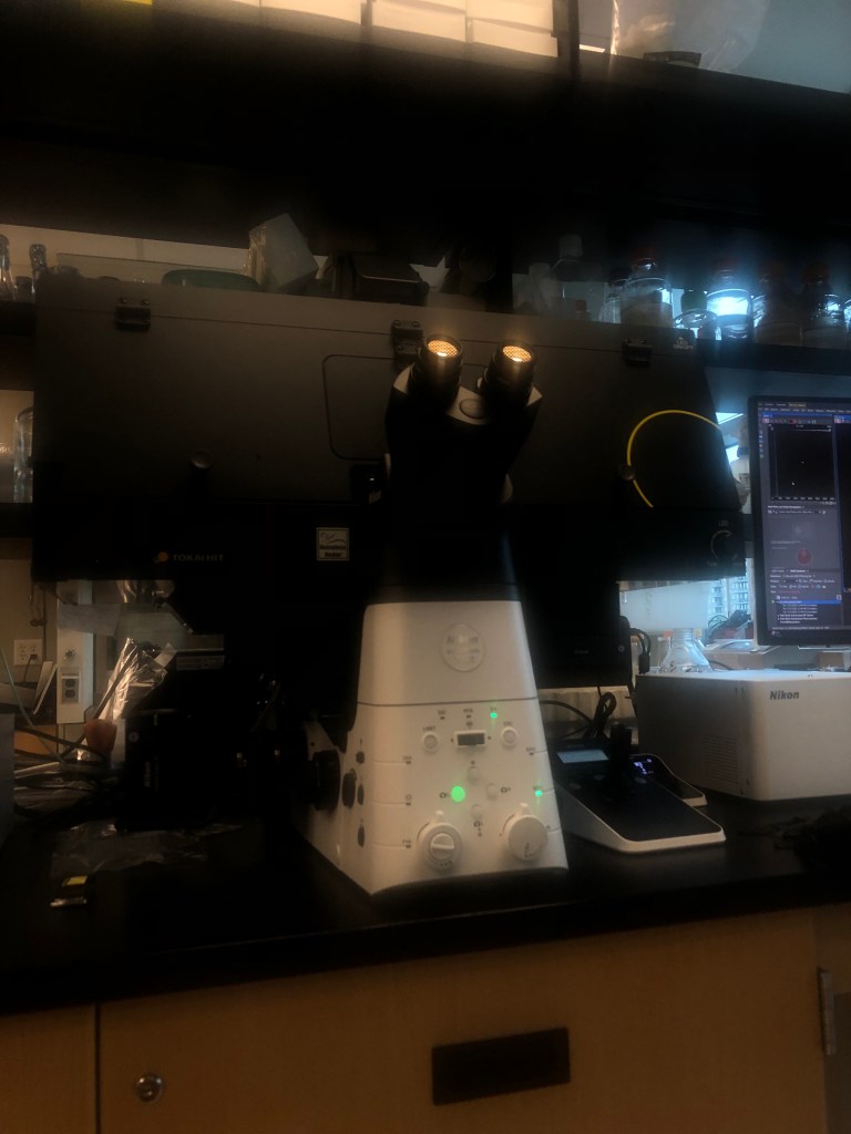
The next stop was the microscope and computer they use to evaluate the cells once inside the fish to see if the cancer has grown, remained the same, or shrunk. If you look at the attached picture, you can see the outline of the fish and the glowing red part is the cancer cells. The new microscope allows them to take photos automatically to better monitor the state of the cells. A bioluminescent substance is used for creating a transgenic zebrafish line (a zebrafish that expresses a specific “human” mutation or other). For transplantation of cancer cells in larval zebrafish a fluorescent dye is used to identify the different cells.
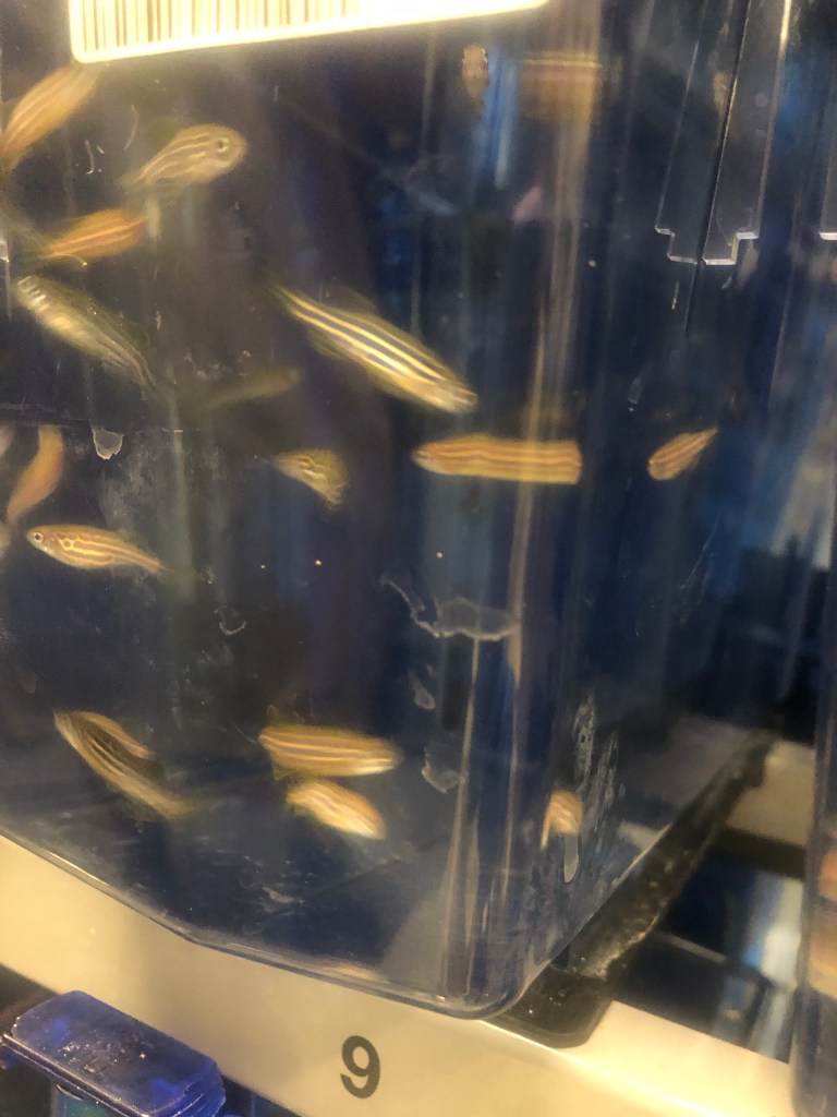
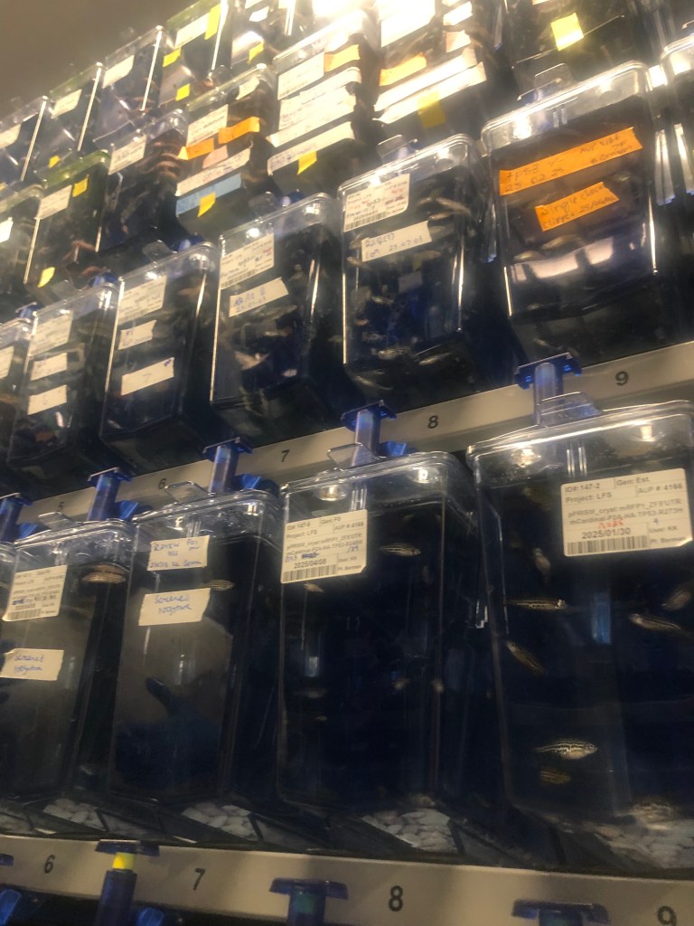
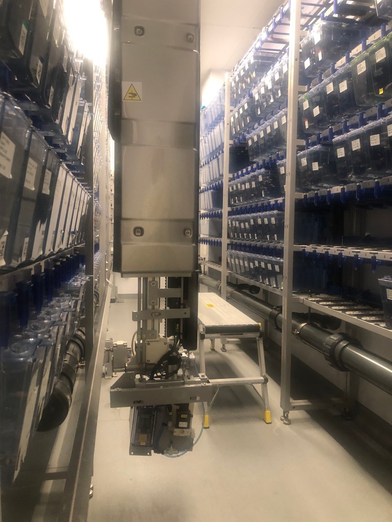
The final part of the tour was probably my favourite. It was time to go to the fish room. It is in the basement so that the light can be controlled to regulate the cycles of the fish. Also, the water is very heavy so it makes sense to have it on the lower floor. A few short elevator rides later, we donned yellow gowns (hello flashback to isolation) to prevent any foreign bacteria from entering the fish room and compromising the zebrafish. Before entering the room, there was a sterilization mat for shoes, but for my chair they sprayed it with the same disinfectant. Once again there was an option for those in a chair. Once inside I didn’t know where to look, there were so many fish! These fish had not been injected with any cancer cells, as they are used for breeding. Their “children” become the larval that are used in the program. I was shown the different fish, their automatic feeding machine, and the breeding tank. So cool to think how some of these fish could be a part of changing history and the way we treat childhood cancer.
This tour was such an amazing experience. It makes me even more excited to be a part of this project. The work they are doing is so important and will save many lives. Personalized treatment could be the future of childhood cancer treatment and I feel honored to be even a small part of it. I will continue to lend my expertise as long as they will have me. I loved how accessible the lab was for me to navigate and how interested the team was in what I had to say. A special thank you goes to Jason Berman for opening his lab up to me, and to Nadine for facilitating this tour. I can’t wait to see what the future holds!


Leave a comment